From most of the works I have read on medical image segmentation, researchers first aim to interactively manually segment training data sets. These segmented data sets are then possibly fed into a machine learning process whereby a model is produced. This model is then capable of recognizing unseen instances of its shape, thus fitting itself to the new instance. Fitting here means segmentation, since the model has fitted itself and segmented an instance.
This all sounds ok, but the manual interactive segmentation part leaves us thinking: is it that easy to manually segment all those training sets by hand?? That's the reason why we are trying so hard to create ways of automatically segment, since manual segmentation is so strenuous and time consuming. Although, it depends on the sort of segmentation tool you are using and your experience in segmenting that particular structure; this is definitely not trivial.
I think I would be wrong saying that manual segmentation is always difficult. For some structures it can be relatively easy, and in some cases quite trivial. For the left atrium, it's a nightmare. The main reason being: there are lots of other blood vessels or arteries which run over (for example) the PV drainages of the left atrium. The diagram below illustrates this:
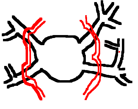 This is a poorly hand-drawn mspaint illustration. The main point I want to bring out are the red vessels which run over the black-coloured bordererd left atrium and its PV drainages. If one were to use a segmentation tool which enabled the user to pick a seed point (starting point of the region-growing segmentation process), and define a bounding box that defined the boundaries of the region-growing process; it would be impossible to segment out the left atrium in one go. A careful and interactive segmentation is required, possibly taking a great deal of time, depending on experience and the segmentation tool's capabilities.
A technique which I tend to follow is to first start out with the core of the atrium. Once the core is segmented, I move out to the PV drainages, segmenting each PV drainage one at a time. This also requires me to define bounding boxes for each of these drainages, and possibly also sometimes changing the threshold levels. This is since, the blood intensity at the drainages tend to be a lot less (35-150) than that of the core of the atrium (typically 60-150). Before getting a properly segmented atrium, it takes on an average 10-15 steps of stichting (union of segmented results) and removing unwanted regions.
After a visit to the cardiologist, I learned that this is exactly what cardiologists do these days. They strenuously join pieces of the left atrium from a 3D rendering of an MRI scan. There seems to be an urgent need of an automatic segmentation tool for the left atrium.
No comments:
Post a Comment