Friday, November 17, 2006

Surface and volume renderings of Left-atrium MRA

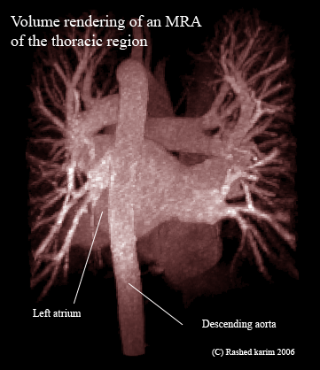
We are ready to show our results and get it verified from the cardiologist. Here are some images showing both volume and surface renderings of the Left atrium with respect to the entire thoracic region. These are MRA scans where the patient is injected with a radio-contrasting agent such as Gadolinium, hence causing the blood vessels to appear as bright structures.


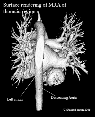
Following is a surface rendering of the above image. One can see the surrounding structures and the descending aorta.


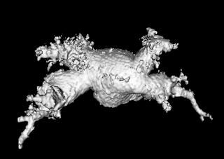
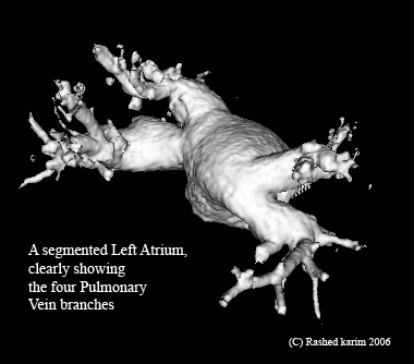
The following image is a surface-rendering of the left atrium which was posted earlier. The previous post featured the same image, however with a lot of noise generating from the extra triangles which were produced as a result of choosing an incorrect threshold in the marching cubes algorithm.


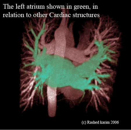
This is apparently one of cardiologists' favorites. The following is a volume rendering of the left atrium and can be seen segmented in green and viewed w.r.t. to its neighboring structures.

Monday, November 13, 2006

A second Atrium

Finally I have got my hands on a second atrium MRA. It is quite different in shape and topology, from the first atrium. It is quite intriguing how such varying shapes of the same organ can occur in nature. The atrium is much larger in shape compared to the previous one, and has thicker PV branches. Such topological variations can make it very difficult to model the shapes using statistical models. One might need to resort to incorporating other techniques that can encode a-priori information, such as probabilisitic atlases.


The MRA data appears to contain a significant amount of noise. I am not entirely certain if this is due to actual noise, or if it's being caused by the rendering algorithm. In the near future, I will be looking more closely into this patient's MRA data and attempt to acquire better images of the segmentation.

Friday, November 10, 2006

Left atrial shape changes

I have come across scores of works which investigate the ventricular volume changes during the cardiac cycle. It has important applications, and is primarily used to quantitatively assess functional parameters such as wall motion, wall thickness and ejection fraction. Considering the fact that the ventricle changes shape, it is not unusual to expect the left atrium to also change its shape. The systole (when the heart contracts) and diastole (when the heart relaxes) are evident in all the four components of the heart. Little work has been done to investigate the atrial volume, and the only piece of work I could find is this. The authors investigate the changes in left-atrial volume in healthy children using 3D Echocardiography.

The changes in left atrial volume can have implications in RF ablation surgery. H. Zhong et. al. describes the RF surgery well in here by comparing it to a painter trying to paint a wall (left atrial endocardium) with his brush (catheter). Assuming that the painter has made an impression of what he is going to paint before hand, on a piece of paper. If the wall started moving, it would be quite trivial to paint it if the painter's motion and the wall's motion are registered (finding a one-to-one correspondence) , along with that of his impression. The hurdle is to be able to solve this correspondence problem and register our 4D model of the atrium with that of the acquired MRI/CTs. H. Zhong et. al. attempts to solve this in their work.

The MRI datasets came in just today, and I will be looking at more atrium segmentation (for training data) in the coming week.