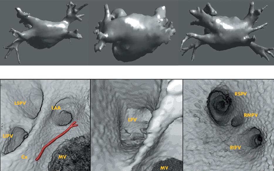
The left atrium, like many organs of the body, comes in different sizes and shapes. They exhibit quite a wide range of differing morphologies. After reading through a medical journal paper by J. Sra et. al. I started to realize how complicated left atrial segmentation can get, due to its varying morphology. Sra's paper looks at 3D reconstructions of left atriums and down below is an image of three varying topologies:
The image on top is the 3D rendering of the left atriums, and the images below are the corresponding endocardial views. If we look the left and right pulmonary veins, we notice that there is a great degree of variation. The far-most left atrial image is what I would call the standard atrial configuration. The middle atrial image is what surprised me the most, and something I have never come across. The right atrial image is also unusual since it has a right middle pulmonary vein portruding out of the right panel.
The paper talks very little about segmentation, and seems more like a review of registration technique and how both segmentation and registration can benefit the medical community, especially in the treatment of cardiac arrhythmias. Total automated segmentation of the left atrium is still far from becoming a reality, and most segmentation techniques I have come across yet requires a great deal of user interaction.
No comments:
Post a Comment