After doing a thorough google search, it was difficult for me to locate an MRI showing the left and the right atriums, together with the ventricles. I am posting some MRI slices obtained by subtracting pre and post-angiographs to remove bone structures. What you see in these MRIs is blood travelling through the different vessels and heart structures. 'Post-angiograph' is after the blood is injected with Gadolinium (Radiocontrasting agent) and what you see in the MRI here are the bright regions indicating the blood travelling through the different structures. However, a pre-angiograph of the thorax region shows nothing really, and only the bone structures.
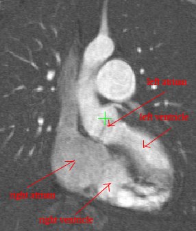
Above is an MRI showing the heart with a long-axis view. But beware! What you see is only the blood travelling through the different regions of the heart, and the radiocontrasting agent in the blood making it appear bright in the MRI.
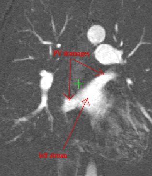
Above, is a better picture of the left atrium together with the PV drainages. There are actually more drainages to this atrium, and these appear in other slices. The left atrium is situated right next to the right atrium and extends to behind the right atrium.
No comments:
Post a Comment