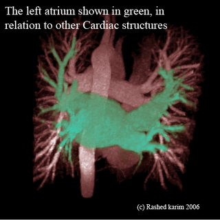
We are ready to show our results and get it verified from the cardiologist. Here are some images showing both volume and surface renderings of the Left atrium with respect to the entire thoracic region. These are MRA scans where the patient is injected with a radio-contrasting agent such as Gadolinium, hence causing the blood vessels to appear as bright structures.

Following is a surface rendering of the above image. One can see the surrounding structures and the descending aorta.

The following image is a surface-rendering of the left atrium which was posted earlier. The previous post featured the same image, however with a lot of noise generating from the extra triangles which were produced as a result of choosing an incorrect threshold in the marching cubes algorithm.

This is apparently one of cardiologists' favorites. The following is a volume rendering of the left atrium and can be seen segmented in green and viewed w.r.t. to its neighboring structures.

No comments:
Post a Comment