I have been experimenting to find out how my implementation performed in segmenting the atrium. Here is one of the cases I have been looking at very closely. I especially like this case since the original image from which the left atrium needs to be extracted is quite 'mesh'-ed within the MRI with the atrium concealed completely inside. Here's the original image:
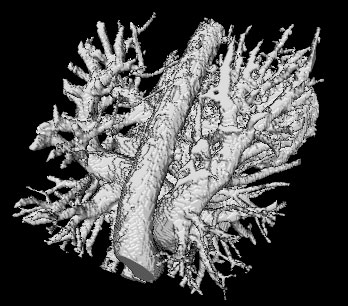
This is almost the entire thoraic region segmented from an image which was obtained by subtracting a post-angiographed from a pre-angiographed MRI (in order to remove the bone structures).
The atrium, after computing the local maximas and basic components surprisingly yielded very few orphans (those which dont belong to a basic component). This is however, not the case with some other segmented images/MRI. I quite do not understand yet what feature of an Euclidean distance transformed image causes these orphans to occur. However, a very preliminary speculation has led to me thinking that it might be because of the absence of local-maximum points in the vicinity of image-edges. Usually, it is the case that when searching for a voxel's basic component close to an edge of the image, the path gets led to a neighborhood of voxels with 0 EDT values. Once it enters such a neighborhood, the search terminates, since the path has exited the EDT-transformed image and it's impossible to search any further. There could be ways of somehow searching further by querying perhaps the original image itself. I might be soon looking at these possibilites soon.
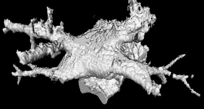
Here is a segmented atrium I obtained by running the implementation on the original image above. The good thing about this segmented atrium is that it reveals all the pulmonary-vein drainages, in addition to also showing how the drainages are branching out.

The atrium, after computing the local maximas and basic components surprisingly yielded very few orphans (those which dont belong to a basic component). This is however, not the case with some other segmented images/MRI. I quite do not understand yet what feature of an Euclidean distance transformed image causes these orphans to occur. However, a very preliminary speculation has led to me thinking that it might be because of the absence of local-maximum points in the vicinity of image-edges. Usually, it is the case that when searching for a voxel's basic component close to an edge of the image, the path gets led to a neighborhood of voxels with 0 EDT values. Once it enters such a neighborhood, the search terminates, since the path has exited the EDT-transformed image and it's impossible to search any further. There could be ways of somehow searching further by querying perhaps the original image itself. I might be soon looking at these possibilites soon.
Here is a segmented atrium I obtained by running the implementation on the original image above. The good thing about this segmented atrium is that it reveals all the pulmonary-vein drainages, in addition to also showing how the drainages are branching out.
No comments:
Post a Comment