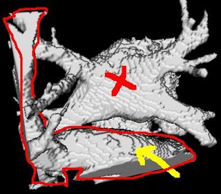
Segmentation of the left-atrium will usually yield connecting extra-cardiac structures. These could be parts of the ventricle and other vessels nearby (such as the aorta). Here is an image of what an initial segmentation would produce:

The structure crossed in red, is the left atrium. The red-outlined structure is presumably the aorta passing right infront of the left-atrium, which was segmented along with the left atrium, in the process of segmenting some of the atrial pulmonary vein drainage endings.
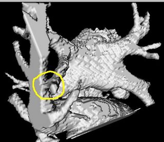
The cardiac structure attached to the left atrium was observed to be attached at points where the blood pool narrows down and opens up into the structure. However, it was also observed to be attached at points which are not narrowed blood pools. The image below is an attachment which is narrowed:

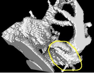
And the image below shows an attachment which is not narrowed.The cardiac structure attached to the left atrium was observed to be attached at points where the blood pool narrows down and opens up into the structure. However, it was also observed to be attached at points which are not narrowed blood pools. The image below is an attachment which is narrowed:


This makes the removal of such post-segmentation artifacts non-trivial, although there is still a possibility of removing most of these by detecting blood-pool narrowings (M. John et. al. MICCAI 2005).
No comments:
Post a Comment